
Raman Spectroscopy
Why?
Early disease detection is a primary requirement for a successful treatment of any disease.
In this context the detection of a disease indicator in body liquids (providing an easily accessible fairly non-invasive sample) will be beneficial since it could spare some of the risky tissue biopsies and offer the potential to detect cancers very early - even before the onset of symptoms - with just a few drops of body liquid (predominantly blood).
Raman based technologies (e.g., resonance Raman, SERS, CARS, SRS etc.), in combination with innovative chemometric approaches and innovative fibres to bring laser light to the point of interest (e.g. hollow organs) and to collect the Raman scattered light, have shown their great impact on biomedical research since a Raman spectrum provides a molecular fingerprint of any biological sample.


What we do in PHAST:
The following directions will be pursued:
1.
Study and development of novel SERS-based optofluidic devices, particularly biological protocols consisting in surface functionalization of metal-dielectric nanostructures for the detection and quantification of tumour biomarkers aimed to perform early cancer diagnostics via a liquid biopsy approach.
This activity merges technologies devoted to synthesis of innovative SERS nanostructures and/or labels and surface biofunctionalization as well as novel microfluidic devices.
2.
Study and development of Standard Operating Procedures (SOP) for point-of-care RS (i.e. used on patient bedside) to detect cancer markers in body liquids (e.g. blood, urine, pericardium liquid etc.).
Here the biggest challenges lie on the systematic evaluation of reproducible Raman sampling methods (e.g. liquids in microcuvettes, capillaries, or after injection in microfluidic chips). This liquid biopsy approach will open new, cheaper and less intrusive ways to diagnose cancer or assess how cancer responds to treatment.
ESRs involved:
ESR 1
Integrated liquid biopsy for cancer diagnostics via genomic markers detection
ESR 2
Point-of-care Raman microspectroscopy for detecting tumour markers in body liquids
Diffuse Optics
WHY?
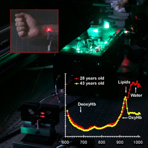
Among other clinical diagnostics modalities, Diffuse Optics - the study of photon migration through highly scattering media - has the peculiarity to convey chemical (i.e., composition), functional (e.g., oxygenation, blood flow), and structural (fibre and cells arrangements) information from depths of few cm within the tissue.
Due to its non-invasiveness, it has raised great interest for diagnosis and monitoring of breast and brain functional status and pathologies.


WHAT WE DO IN PHAST
Three directions will be pursued.
1.
the adoption of novel time-gated approaches which will permit to reach internal organs via single-fibre fine-needle optical biopsies;
2.
the incorporation of DO information into multimodalities probes (e.g., imaging, fluorescence) where DO will complement morphological info with functional and chemical monitoring for theranostics interventions with real time classification of suspect lesions;
3.
Monitoring cancer treatment, in particular PDT via alterations in tissue absorption, and neoadjuvant chemotherapy via changes in breast lesion composition and structure.
Besides exploiting novel photonics devices (e.g., fast gated single-photon detectors, compact laser sources, microelectronics processing of optical signals) research will open new applicative directions, particularly towards theranostics and therapy monitoring.
Strong experience in performance assessment and standardization will be shared for high quality results and smoother industrial deployment.
ESRs involved:
ESR 5
Hybrid diffuse optical monitoring and theranostics with blood flow and oxygen metabolism biomarkers on pre-clinical models
ESR 6
Time-gated diffuse optical spectroscopy for deep tissue diagnostics
ESR 12
Development of advanced online dosimetry algorithms for photodynamic therapy of tumours
ESR 13
Diffuse optical monitoring and prediction of neoadjuvant chemotherapy for personalized breast cancer management
ESR 14
Tissue identification for surgical guidance using biophotonics
Multifunctional Fibers
WHY?
Current optical fibre sensors for healthcare are mainly used as light delivering devices for endoscopes. Only recently, optical fibres have included smart functionalities like for example fibre Bragg grating for 3D shape sensing or the use of different materials apart from the glass of which they are made to integrate e.g. along the fibre optical sensors or piezo microactuators.
The application of these novel technologies towards the realization of multifunctional sensors/actuators is still a green field especially in the biomedical sector.


WHAT WE DO IN PHAST
Two studies will be carried out involving the following activities:
1.
Study and development of novel distributed fibre-based pressure sensor for the aid in the diagnosis of angiogenesis in cancer (brain tumours) and monitoring. The research will aim at optical sensors designed to be embedded in endoscopes/arterial catheters for intravital monitor and diagnostics and their validation in phantoms.
2.
Development of an optical probe able to combine several functionalities in the field of cancer diagnostics and treatment, thanks also to the inclusion, directly into the fibre, of materials which are traditionally not used in fibre optics.
The final goal is to integrate advanced functionalities like in-situ drug release, light delivery for treatment (e.g., photodynamic therapy, photothermal therapy), electrical signal transport (e.g., RF ablation and sensing) and diagnostics (e.g., time-domain diffuse optical spectroscopy) are foreseen.
ESRs involved:
ESR 4
Integrated optical fibre sensors for intravital monitoring
ESR 11
Towards multifunctional multimaterial optical fibres for diagnostic and treatment
Multimodal Imaging
WHY?
Biophotonic imaging methods as stand-alone techniques are typically highlighting only an aspect of pathological alterations. To improve the diagnostic potential, it has been shown that the combination of different spectroscopic imaging methods (e.g., Optical Coherence Tomography - OCT, SHG, TPEF, CARS, SRS, Raman etc.) into a multimodal approach is very beneficial in providing conceptually novel insights into complex biological media.


WHAT WE DO IN PHAST
The following multimodal imaging approaches will be pursued:
1.
Multimodal imaging utilizing imaging approaches with similar image acquisition times. Here modalities requiring the same or similar experimental equipment should be combined such as the integration of various nonlinear techniques (SHG, TPEF, CARS, SRS etc.).
2.
Imaging techniques yielding a large field of view of morphological information (e.g., OCT) will be combined with molecular specific approaches (e.g., Raman, CARS, TPEF) to link morphological information with a richness of molecular detail of selected points or confined areas.
PHAST will systematically evaluate various multimodal microscopic and endomicroscopic strategies with respect to achieving a real-time diagnosis of various cancer pathologies. This involves the combination of new laser technologies, with novel micro-optic imaging systems (based on gradient-index micro-lenses) and advanced algorithms for real time image analysis.
The transformation from a multimodal surgery microscope to a multimodal endoscope involves innovative fibre concepts, which will be also researched.
The specific detection of malignant tissue during curative surgery is the most important precondition for complete tumour removal. Therefore, multimodal imaging will be further extended by selective laser tissue ablation.
ESRs involved:
ESR 3
Spectral tissue imaging for ex-vivo cancer diagnosis and survey
ESR 7
Multimodal nonlinear imaging for clinical diagnosis in combination with laser tissue ablation for selective tissue removal
ESR 8
Micro-optical imaging system for multimodal non-linear endospectroscopy
ESR 9
Multimodal endo-microscopy for improved in-vivo colorectal cancer diagnosis
ESR 10
Multimodal intraoperative handheld forward-imaging probe
Stay in touch
subscribe to our newsletter service
